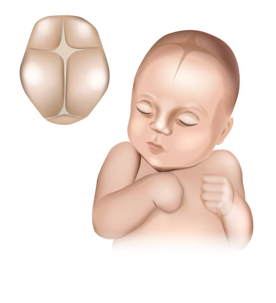
An illustration of a realistic babys head showing the fontanelles present at birth. Cranial sutures and fontanels. Baby s Soft Spots.
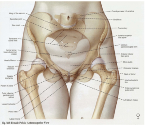
The fetal skull refers to the bony framework of the unborn baby’s head. Compared to an adult, a fetus has a relatively large head in proportion to its body. This size difference is even more pronounced when compared to the mother’s pelvis, making the fetal skull the largest and most significant part of the baby to pass through the birth canal during labor. For a smooth and complication-free birth, the skull must adjust to fit through the pelvic opening.
Why It Matters for Mothers and Midwives
-
Helps anticipate delivery outcomes—whether labor may be prolonged, difficult, or uncomplicated.
-
Aids in deciding the safest delivery method—natural (vaginal) or surgical (caesarean section
-
It tells the presentation of the baby and can detect abnormal presentation
- It is the most important part of the fetus as it contains the most delicate organ. the brain.
Developmental Timeline
The fetal skull begins forming early in embryonic development, with ossification centers appearing around the 8th week of gestation (pregnacy). Unlike the adult skull, which consists of fused plates, the fetal skull contains multiple unfused bones connected by fibrous joints called sutures and fontanelles (soft spots). This design allows for:
- Brain growth accommodation during fetal development
- Skull compression during delivery (molding)
- Continued brain expansion after birth
Divisions of the fetal skull
The fetal skull is made up of three main areas — the dome-like vault, the sturdy base, and the face, each with a unique function during birth.
The vault
Main Divisions of the Skull
The fetal skull is divided into three major sections:

-
The vault is the top, rounded part of the skull that goes from the eyebrows to the back of the head. The bones here are thin and a bit flexible, so if a lot of pressure is applied, it can hurt the soft brain underneath.
The base is the bottom part of the skull. It’s made of strong, tightly connected bones that protect important parts of the brain, especially in the area called the medulla oblongata.
The face is made up of 14 small bones that are tightly joined together and cannot be squeezed. It starts from the bones around the eyes (orbital ridge) and goes down to where the chin (mentum) connects with the neck.
The bones of the vault
The bones of the vault (skull) start forming in soft tissue called membrane. They harden from the center outward in a process called ossification. At birth, this process isn’t complete, so there are small gaps between the bones called sutures and fontanelles. Each bone has a raised area where the hardening begins (protuberance). The skull doesn’t fully harden until early adulthood.
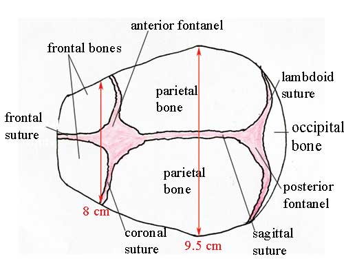
The bones of the vault consist of:
-
Occipital Bone – at the back of the head; includes the foramen magnum, where the spinal cord exits.
-
Parietal Bones (2) – located on each side of the head; ossification starts at the parietal eminence.
-
Frontal Bones (2) – form the forehead, known as the sinciput; they begin as two bones that fuse into one by around 8 years old. The ossification centre of each bone is the frontal eminence.
-
Temporal Bones (upper portion) – found on both sides of the head and partially contribute to the vault.
Another important part of the fetal skull is the SUTURE AND FRONTANELLE

Sutures: The sutures are the cranial joints formed where two bones meet.
These sutures allow the skull bones to overlap slightly during birth and separate to accommodate brain growth after birth.
These fibrous connections between skull bones include:
- Sagittal suture: Runs from front to back along the midline (between the two parietal
bones.) - Coronal suture: Runs across the top of the head from ear to ear (separates the frontal bones from
the parietal) - Lambdoidal suture: Curves across the back of the head (separates the occipital bone
from the two parietal bones.) - Frontal suture: it lies between the two halves of frontal bones.
Fontanelles: The Important “Soft Spots”

Those famous “soft spots” on a baby’s head are called fontanelles (pronounced: fon-tuh-NELS). These aren’t dangerous open areas—they’re covered with tough protective membranes. When you gently touch them, you might feel:
- A slight pulse (just the normal heartbeat)
- A slight inward or outward curve (depending on the baby’s position and hydration)
The largest soft spot sits on top of the head and typically closes between 9-18 months as the puzzle pieces of the skull grow together. You could also say it’s a place where two or more sutures meet,
These membrane-covered spaces between skull bones serve crucial purposes and are divided into two:
- Anterior fontanelle (Bregma): Diamond-shaped, located at the top of the head where four bones meet (Juction of the saggital coronal and frontal sutures) . Typically, 4-6 cm at birth and closes between 18 months. if felt during a vagina examination, it indicates that the head is not well postioned.
- Posterior fontanelle (Lambda): Smaller, triangular-shaped, located at the back of the head. Usually closes by 6 weeks after birth. it felt during vaginal delivery, it indicates that the fetus if presented properly.
As a parent, you can safely touch these areas—they’re protected by tough membranes. Your midwife may assess these spots during antenatal and postnatal checks.
The sutures and fontanelles described below permit a degree of overlapping of the skull bones during labour, which is known as moulding.
landmarks of the fetal skull
This is also an important area as it will give you insight into understanding proper positioning of the fetus.
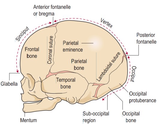
- The occiput region – the area occupied by the occipital bone
- The vertex region – highest point in fetal skull (bounded by the posterior fontanelle, two parietal eminence and anterior fontanelle)
- Sinciput – Forehead
- Mentum – Chin
- Glabella – bridge of the nose or point below the eyebrow
- suboccipital region – the part below the occipital protuberance
- face – extend from the obital ridges and root of the nose to the chin (mentum)
The Birth Process: How the Skull Adapts
During labor and birth, we observe:
Molding
The baby’s head temporarily changes shape as it passes through the pelvis. This process, called “molding,” involves the skull bones sliding slightly over each other to reduce the diameter of the head.
The degree of molding depends on:
- Duration of labor
- Position of the baby
- Shape of the mother’s pelvis
- Whether it’s a first or subsequent birth
Most molding resolves within 24-72 hours after birth—nature’s way of returning the head to its intended shape.
Engagement and Descent
The fetal head typically engages in the pelvis in an asynclitic position (slightly tilted) and rotates during descent to maximize the fit between the head and the pelvis. The sutures and fontanelles allow this crucial adaptation.
After Birth: What Parents Should Know
Normal Findings
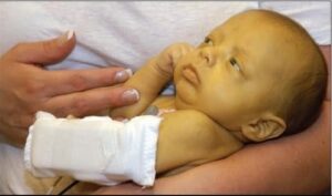
- Caput succedaneum: A diffuse swelling of the scalp that crosses suture lines; typically resolves within days
- Cephalohematoma: A collection of blood between a skull bone and its covering; does not cross suture lines and may take weeks to resolve
- Overlapping sutures: Edges of bones may feel slightly raised; typically resolve within days
When to Contact Your Midwife
Reach out if you notice:
- Excessive bulging or depression of fontanelles
- Premature closing of sutures (causing unusual head shape)
- Rapidly increasing head circumference
For Student Midwives: Clinical Skills
Learn to:
- Identify fontanelles and sutures during vaginal examinations
- Assess fetal position using skull landmarks (fontanelles and sutures)
- Recognize normal variations versus potential complications
- Measure head circumference accurately
The Wisdom of the Body
As midwives, we honor this remarkable design that allows babies to navigate the journey of birth. The fetal skull exemplifies the body’s inherent wisdom—flexible enough for birth, yet protective of the precious developing brain.
Understanding these normal processes helps parents feel confident about their baby’s development and helps students appreciate the intricate dance between mother and baby during birth.